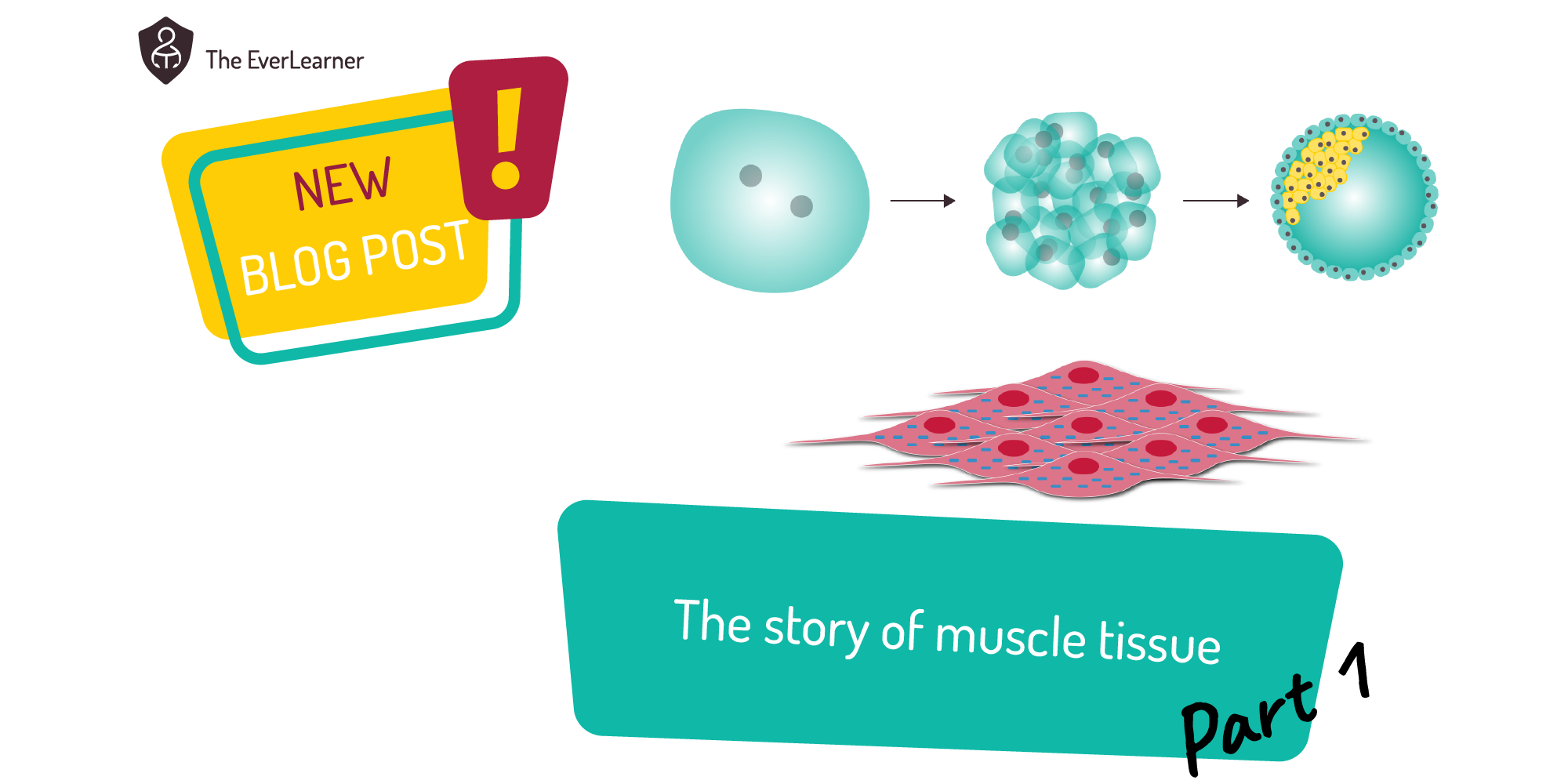
%20Text%20(Violet).png)
Welcome back to the story of muscle tissue. If you have already read Part 1, you will know that today’s Part 2’s post is all about muscle structure and function.
The structure and function of muscle tissue
Muscle tissue, as established in Part 1 is a highly specialised collection of muscle cells that is capable of generating force. There are three types of muscle tissue in the human body:

I am going to focus on the structure and function of skeletal muscle.
Skeletal muscles are precisely structured to perform their role. The key details of skeletal muscles are as follows:
- They are connected to bone at both ends via tendon tissue.
- There is a moving end known as the insertion and a static end known as the origin.
- They apply pulling force in one specific direction only.
- The force they apply has the tendency to move the bone onto which it inserts.
Like this:

And this: 
In the case shown above, we can see only the insertion end of the muscle. We can clearly see the tendon tissue sitting between the skeletal muscle and the long bone and, once again, this tendon tissue will transmit force from the muscle tissue to the bone tissue causing it, potentially, to move. A little note of detail may help here. BOTH the muscle tissue and the tendon tissue are surrounded by the same connective tissue or sheath. This is called the epimysium and it helps to surround both tissues and to prevent the force generated by the muscle tissue from being lost or dissipated. When athletes injure their tendons or muscles more superficially, it is often this connective tissue that is damaged and, rather than the muscle or tendon tissue being damaged directly, the epimysium is split or “nicked” and this can lead to a loss of force as well as pain.
But if we direct our attention to the right of the image, another piece of intuition can grab us or our students:
The muscle tissue is structured with units and further subunits all of which have a long, thin profile. Notice the yellow fascicle, for example. A fascicle is a bundle of muscle fibres, all of the same nature (fibre type… see below) surrounded by connective tissue, this time called the perimysium. These fascicles are functional units. This means that they can work (pull on the bone) whether or not other fascicles around them are doing likewise. This is one way in which human beings can produce more and less powerful muscle contractions. Different units within the muscle tissue can fire or not. Imagine, for example, the rectus femoris (quadriceps group) of a weightlifter attempting a world-record deadlift. All of the units of the muscle tissue would fire and a maximal, or close to it, contraction would occur.
Perhaps even more interesting is that these subunits can be “rested” during ongoing physical activity. Consider an Olympic kayaker, for example: as they repeatedly pull their paddle against the water, the muscle tissue is “smart” enough to switch off units that are fatigued, assuming that the muscle tissue is not nearing maximal force. This is known as spatial summation. In other words, the sum of the subunit force contraction is spaced out, allowing parts of the muscle tissue to recover. Do your students know this? If so, how can they use their knowledge in their PE lessons, extracurricular or sport performances?
Going further
Many of you will know that the microscopic structure of muscle tissue does not stop at a muscle fibre. Absolutely not. If I were to attempt to ruin the image I have been using until now, I might make an edit something like this:

Notice the pink filaments exiting the muscle fibre. These filaments are made from protein and are the force-producing units of muscle fibre cells.
If we were to look very closely, via a light or, even better, an electron microscope, we might expect to see structures like this:

The muscle fibre has overlapping protein filaments of different types. The filament illustrated in blue and yellow is called actin and the red and pink filament with the little hockey stick attachments is called myosin. It is these fibres pulling against one another, in one specific direction, that actually generate the muscle’s force. Sometimes I like to think that, in order to apply force and create contraction, the myosin (heads) have to walk along the actin filament. As you would imagine, there’s a little more to it than this but the intuition of what I have described is reliable.
Teaching on the edge… accidentally!
I’m slightly nervous about what I’m about to write below. The thing is, when I first taught this to students, I had no idea that the topic of sliding filament theory can be very susceptible to a double entendre. I remember when I was a 2nd year teacher and I taught this to my A-level group in Oxfordshire, the students all started giggling. To me, it was just the microscopic muscle structure, and I had no intent to be “naughty”. It went something like this:

Looking back now, I can see why the giggling occurred. In subsequent years, I have appropriately made the most of this, just flirting with the suggestive nature of the double entendre and, somehow, it has turned a confusing, terminology-based concept into a memorable experience for students. Clearly, with some groups, there have been times where I have actively avoided this possibility based on the character of the group, or sensitivities, or just being unsure of the response.
So, I will choose to leave Part 2 at this point. I hope you find this update interesting and I really hope it has encouraged you to discuss muscle tissue with your groups in a refined and accurate way.
Come back for Part 3 which will drop very soon and will contain topics such as antagonistic pairs, muscle contraction types, fibre types and much more.
James